
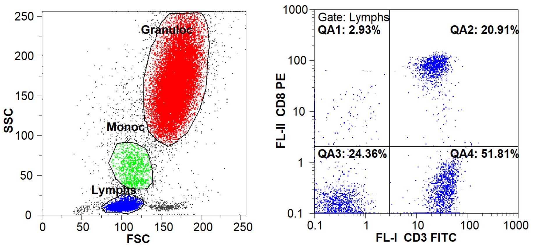
Immunophenotyping is one of the most commonly used applications for flow cytometry. Single cell, multicolor fluorescence analysis of blood and other biological tissues identifies and quantitates the different types of cells for research and medical purposes. Sample preparation involves the incubation of the cells with fluorescently tagged monoclonal antibodies that specifically bind to different cell types. The fluorescent dyes then fluoresce when excited by the appropriate lasers of the flow cytometer. Using the forward scatter (FSC) and side scatter (SSC) of the laser light, cells can be identified by their physical properties such as size and density. Subsets of cells are then classified and quantified based on the light scatter and fluorescence for each cell.
Among the many uses for immunophenotyping, one of the most useful is the identification of the type of cells that have become cancerous in leukemia. By identifying which type of white blood cell has become malignant in leukemia, a treatment can be selected that is most effective against that type of leukemia. The figure above shows how light scatter can be used to distinguish the lymphocytes from the monocytes and granulocytes and fluorescence can be used to identify and quantitate the lymphocytes that are T lymphocytes (CD3-positive) and which of those are T Suppressor/Cytotoxic lymphocytes (CD3-positive and CD8-positive).